The Team approach
The Team Approach
At SCARS Center we use a team approach to cure skin cancer and provide cosmetically conscious results in one, convenient location.
Skin Cancer Facts & Figures
Facts & Figures
SCARS Center surgical specialists encourage patients to know the facts about skin cancer. Protect yourself and your loved ones by learning more about skin cancer today.
Skin Cancer Prevention
Skin Cancer Prevention
Learn the risk factors, warning signs, and behavioral patterns of skin cancer as your first line of defense against this potentially life threatening disease.
Skin Cancer Aftercare
Skin Cancer Aftercare
Our physicians place a special emphasis on skin healing and scar management techniques so patients can feel confident in their appearance after recovery.
Half a Facelift? Full Life Improvement!!
Frank Ware’s journey with the SCARS Center and Dr. Madorsky highlights the synergy between reconstruction and aesthetics. After a melanoma removal on his left cheek, he underwent a specialized repair of his right cheek—decades after facial paralysis following brain surgery. A deep plane facelift restored some of the symmetry and function of the right side of his face, enhancing Frank's ability to communicate, his appearance, and his confidence.
King of speed Kenny Bernstein and skin cancer
Kenny Bernstein, known as the King of Speed, is a renowned drag racer and successful buisnessman. In his nearly 40-year career as a racer, he collected dozens of event victories and multiple championship titles, most of these with his iconic Budweiser King Race Team.
In this installment of Skin Cancer Stories, we get to know the King of Speed, talk to him about his illustrious racing career, and explore his journey with skin cancer.
The third and final episode of our new series on Mohs Surgery is now live on Youtube!
This video takes an in-depth look at Mohs Surgery, and how it is performed in the collaborative environment of the Skin Cancer and Reconstructive Surgery Center in Newport Beach.In this episode, we reveal the history of Mohs surgery, and explore the reconstruction process.
The SECOND episode of our new series on Mohs Surgery is now live on Youtube!
This video takes an in-depth look at Mohs Surgery, and how it is performed in the collaborative environment of the Skin Cancer and Reconstructive Surgery Center in Newport Beach. In this episode, we explore common skin cancers, primary risk factors, and how Mohs Surgery can essentially cure skin cancer. We also look at an uncommon case that puts this procedure to the test.
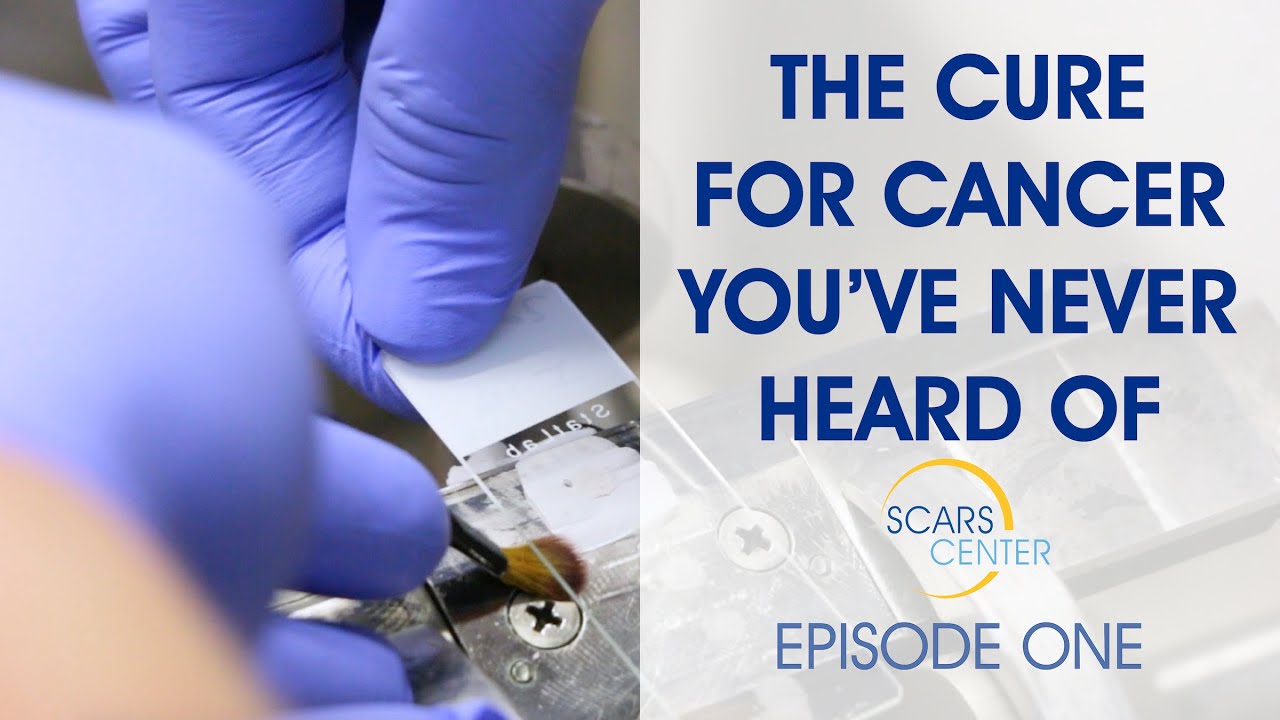
The first episode of our new series on Mohs Surgery is now live on Youtube!
This video takes an in-depth look at Mohs Surgery, and how it is performed in the collaborative environment of the Skin Cancer and Reconstructive Surgery Center in Newport Beach. In this episode, we explore common skin cancers, primary risk factors, and how Mohs Surgery can essentially cure skin cancer. We also look at an uncommon case that puts this procedure to the test.


complete facility for skin cancer management
Skin Cancer and Reconstructive Surgery Center is an all-inclusive skin cancer treatment facility, located in Newport Beach, California. Our center is designed to assist patients seeking a variety of treatment options in one, convenient location.
SKIN CANCER AND RECONSTRUCTIVE SURGERY CENTER OFFERS:
- Medical examination rooms
- A Mohs Laboratory for onsite analysis of skin cancer
- Nonsurgical treatment areas for Superficial Radiotherapy (SRT) and Photodynamic Therapy (PDT)
- An accredited surgery center
- Aftercare center dedicated to scar management, skin care, and facial rejuvenation
CALL NOW TO SCHEDULE YOUR SKIN CANCER EVALUATION
949.719.1800
We have two locations:
180 Newport Center Drive Suite 158, Newport Beach, CA 92660
17451 Bastanchury Rd, Suite 103A, Yorba Linda, CA 92886
MONTHLY SKIN CANCER CONFERENCE
The Skin Cancer and Reconstructive Surgery Foundation is accredited by the Institute for Medical Quality/California Medical Association to provide continuing medical education for physicians.
Skin Cancer Connection Blog
The SCARS CENTER Skin Cancer Connection Blog is the only online collection of skin cancer case studies with discussion and expert analysis for and by physicians.
WHY CHOOSE SCARS CENTER?
SCARS Center is home to some of the most widely respected dermatologists and plastic and reconstructive surgeons in Orange County.
FOLLOW US
STAY UP TO DATE
Receive research updates, inspiring stories, healthy living tips and more.
Orange County's Premier Skin Cancer Treatment Center